Still symptomatic after a concussion? Learn why post-concussion syndrome persists neurologically and what evidence-based treatments actually work.
Most people expect a concussion to resolve within a few weeks. Rest, avoid screens, take it easy, and the brain heals. That is the standard advice, and for many people, it works. But for a significant number of patients, weeks stretch into months, and symptoms like headaches, brain fog, sleep disruption, and mood changes refuse to go away. This is post-concussion syndrome, and understanding why it lingers changes everything about how it should be treated.
The reality is that post concussion symptoms and treatment cannot be approached as a simple waiting game. When symptoms persist beyond six weeks, the brain is not merely healing slowly. Research shows measurable changes in brainwave patterns and neural connectivity that passive rest simply cannot correct on its own. The good news is that targeted, evidence-based interventions exist.
This post breaks down what is actually happening in the brain after a concussion, why the traditional "rest and wait" approach falls short for so many patients, and what active rehabilitation, including qEEG brain mapping and neurofeedback, looks like in practice. If standard advice has not brought relief, this is where to start.
The Six-Week Myth: Why 'Just Rest' Is Not a Treatment Plan
Most people who sustain a concussion are told the same thing: rest, avoid screens, take it easy, and expect to feel better within four to six weeks. For roughly 90% of patients, that advice holds. Symptoms resolve, and life returns to normal.
But a meaningful subset of patients reaches that six-week mark still struggling with headaches, cognitive fog, fatigue, and mood disruption. Many are then told to keep waiting. That guidance has a significant problem: it was established before modern neuroimaging and electrophysiology existed to show what a concussed brain actually looks like during recovery.
The "rest and wait" model was built on observable behavior, not on measurable brain activity. When clinicians had no tools to detect functional disruption at the neural level, watchful waiting was a reasonable default. It is no longer the complete picture.
Why do concussion symptoms linger after scans look normal? The answer points to something the standard recovery model does not account for: post-concussion syndrome is not slow healing. It reflects a nervous system that has shifted into a dysregulated state. Brainwave patterns become abnormal. Communication between brain regions breaks down. These are measurable changes, and passive rest does not provide the targeted input required to correct them.
This distinction matters enormously for patients. Research confirms that mild TBI producing persistent post-concussive symptoms causes lasting effects on cognition, memory, and executive function. Patients still symptomatic at six weeks are not failing to recover. They are experiencing a neurophysiological problem that the rest-based model was never designed to treat.
Recognizing that distinction is the first practical step forward. Recovery from concussion syndrome, for this population, is not a matter of waiting longer. It is a matter of shifting from passive waiting to active, targeted rehabilitation directed at what is actually happening in the brain.
What Is Actually Happening in the Brain After a Concussion
To understand why rest alone cannot resolve persistent symptoms, it helps to understand what a concussion actually does to the brain at the cellular level.
A concussion does not require visible tissue destruction to cause lasting dysfunction. The injury is functional, not structural. Neurons remain physically intact, but the way they communicate and synchronize with each other is disrupted. This distinction matters enormously, because the tools most commonly used to evaluate concussion patients, standard MRI and CT scans, are designed to detect structural damage. They cannot image a communication problem. A scan can appear completely normal while a patient experiences debilitating headaches, cognitive fog, and emotional volatility. This is why so many patients are told "nothing is wrong" and sent home to rest.
The Functional Overlay Model
Research published in the peer-reviewed journal Brain Science in July 2023 formalized this understanding through the Functional Overlay Model, a framework specifically developed to explain why PCS symptoms persist long after the initial injury event. The model proposes two interrelated mechanisms at work.
The first is brainwave dysregulation: measurable abnormalities in the frequency and amplitude of electrical activity across brain regions. These patterns are not abstract. They govern concrete functions including attention, memory retrieval, emotional regulation, and sleep architecture. When the patterns are disrupted, those functions degrade in corresponding ways.
The second mechanism is altered connectivity: the communication pathways between brain regions become desynchronized. The practical consequence is significant. Tasks that previously required minimal cognitive effort, reading a paragraph, following a conversation, managing a schedule, now demand disproportionate neurological resources. This is the direct neurophysiological explanation for the mental exhaustion that characterizes PCS. The brain is not being slow; it is working far harder than it should be to accomplish ordinary tasks.
Why This Changes the Clinical Picture
Reframing PCS as a measurable neurophysiological state rather than a subjective complaint has direct implications for both assessment and treatment. If the problem is functional dysregulation, then the right diagnostic tool must be capable of measuring function, not just structure. Standard imaging was never designed for that purpose. Tools that analyze the brain's electrical activity patterns are better suited to detecting what MRI and CT scans miss. For a more detailed overview of how brain mapping applies in concussion cases, the overview of brain mapping for concussion clients explains this assessment distinction in practical terms.
Equally important: brainwave dysregulation is not self-resolving through inactivity. That is the central problem with passive recovery, which the next section addresses directly.

Why Passive Recovery Fails for Post-Concussion Syndrome
Knowing that PCS involves measurable brainwave dysregulation reframes an important follow-up question: if the problem is neurophysiological, why doesn't rest fix it?
Rest is genuinely useful in the first days after a concussion. It reduces symptom load and gives the brain's acute inflammatory response time to settle. But rest is passive. It removes demand from a dysregulated system without providing the specific input required to correct that dysregulation. Reducing symptoms temporarily is not the same as resolving the underlying pattern producing them.
Neuroplasticity is the mechanism that explains this gap. The brain's capacity to reorganize and restore healthier function is an activity-dependent process. It requires specific, repeated stimulation to drive change. Prolonged rest does not trigger neuroplastic reorganization. It maintains the status quo, and in PCS, the status quo is a dysregulated nervous system.
That matters more with each passing week. When a dysregulated nervous system goes without corrective input, it can consolidate around its maladaptive patterns. Aberrant brainwave activity becomes the brain's new operating baseline, and the longer this continues, the more entrenched that baseline becomes. Passive waiting grows increasingly counterproductive over time rather than simply neutral.
Rest also does nothing for the downstream effects that independently sustain PCS. Research identifies autonomic nervous system dysfunction as central to post-concussion pathophysiology, with abnormal regulation of cerebral blood flow contributing to exercise intolerance and persistent symptoms. Disrupted sleep architecture, chronic autonomic dysregulation, and psychological responses like anxiety and hypervigilance are not side effects waiting to self-resolve; they are active drivers that amplify and extend the symptom cycle. Each requires its own targeted intervention.
The clinical field has responded accordingly. A 2023 interdisciplinary study published in the Annals of Physical and Rehabilitation Medicine documented clinically significant improvements in persistent post-concussion symptoms through a 12-week protocol combining physiotherapy, psychology, and medical intervention. Passive rest was not a comparison arm because it was not considered a viable alternative for that population.
For patients and providers alike, this creates a specific decision point. There is a moment when continued rest stops being conservative clinical management and starts being an obstacle to the active rehabilitation the brain actually needs. Identifying that moment early is one of the most consequential steps in concussion syndrome recovery.
For a closer look at how neurofeedback addresses these neurological patterns specifically, see the benefits of neurofeedback for concussion recovery.
Recognizing Post-Concussion Syndrome: Symptoms That Signal More Than Slow Healing
Knowing that passive recovery has limits is one thing; recognizing that your specific symptoms cross the clinical threshold for post-concussion syndrome is another. The symptom picture matters, because it tells you what kind of help to seek.
Core physical symptoms include persistent headaches, sleep disturbance, fatigue disproportionate to activity level, dizziness, and difficulty concentrating. Cognitive symptoms follow closely: memory lapses, slowed processing, and a "foggy" quality to thinking that makes ordinary tasks feel effortful. Irritability and heightened emotional reactivity round out the most common presentation per Mayo Clinic's documentation of post-concussion syndrome.

Headaches deserve specific attention. Post-concussive headaches are among the most searched symptoms for good reason; they are frequently resistant to standard over-the-counter pain relievers. The reason is mechanistic: these headaches often originate from neurophysiological disruption, including central sensitization and neuroinflammatory processes, rather than the muscular tension or vascular changes that drive typical tension or migraine headaches. Treating a neurophysiological headache with analgesics designed for peripheral causes explains why so many PCS patients report minimal relief.
Sensory sensitivities signal regulatory overload, not adjustment. Light sensitivity (photophobia) and sound sensitivity (phonophobia) indicate that the brain's regulatory systems are struggling to filter and modulate input, not simply adapting to injury. When ordinary environments feel overwhelming, the nervous system is not healing; it is dysregulated.
Emotional symptoms are neurological, not purely reactive. Anxiety, low mood, and social withdrawal are common in PCS, and for many patients their origin is biological rather than a straightforward psychological response to the injury event. The same disrupted neural connectivity that impairs memory and concentration also affects emotional regulation. Depression after concussion or TBI is a clinically documented consequence of functional brain disruption, not simply sadness about being injured.
The clearest clinical signal is symptom clustering that persists beyond six weeks despite following standard rest-based advice. A single lingering headache is different from overlapping headaches, cognitive fog, sleep disruption, and irritability that collectively show no meaningful improvement. That pattern warrants evaluation for active rehabilitation, not more time waiting.
What Evidence-Based Active Rehabilitation Actually Looks Like
Once you recognize that your symptoms are neurologically driven rather than a matter of healing slowly, the natural next question is: what actually works?
Effective rehabilitation for post-concussion syndrome is not a single treatment. It is a protocol-driven sequence of assessment and targeted intervention built around each patient's specific neurophysiological profile. Generic advice cannot fix a problem that looks different in every brain.
Measurement Before Treatment
The starting point is objective data. Clinicians need to identify which brainwave frequencies are dysregulated, in which regions, and how connectivity between those regions has been affected. Without that information, any intervention is essentially a guess. This is the core distinction between precision rehabilitation and the standard "rest and follow up in a few weeks" approach. Measuring the brain's electrical activity before designing a treatment plan is not optional; it is what makes the intervention accurate.
Cognitive Rehabilitation Therapy
Once the neurophysiological picture is clear, cognitive rehabilitation therapy addresses the functions most commonly disrupted in post-concussion syndrome: attention, working memory, processing speed, and executive function. Structured exercises rebuild these capacities progressively, while compensatory strategies help patients manage real-world demands during recovery. This is an established component of post-concussion recovery support and cognitive rehabilitation therapy for TBI more broadly, not an experimental add-on.
Neurofeedback as a Clinically Recognized Modality
Neurofeedback trains the brain to produce healthier electrical patterns by providing real-time feedback on its own activity. The International Society for Neuroregulation (ISNR) published updated clinical guidance on neurofeedback for concussion recovery in April 2026, reflecting a growing professional consensus that this modality belongs in formal concussion syndrome recovery protocols.
Institutional recognition extends further. In April 2024, the WorkSafeBC Evidence-Based Practice Group completed a systematic review evaluating qEEG and neurofeedback for concussion treatment, searching across Cochrane, Medline, and Embase. The fact that a workers' compensation regulatory body formally commissioned that review signals that these interventions are no longer fringe; they are being evaluated at the level of insurance and occupational medicine.
Documentation That Supports Legal and Insurance Claims
For patients pursuing personal injury or workers' compensation cases, this matters beyond clinical recovery. Objective neurophysiological assessments produce timestamped, measurable records of functional impairment. That documentation carries considerably more weight in legal and insurance proceedings than symptom self-report alone, giving affected patients a concrete evidentiary foundation for their claims.
The Role of qEEG Brain Mapping in Post-Concussion Assessment
That objective measurement framework discussed in the previous section depends entirely on the right diagnostic tool. For post-concussion syndrome, that tool is quantitative electroencephalography, or qEEG.
What qEEG Actually Measures
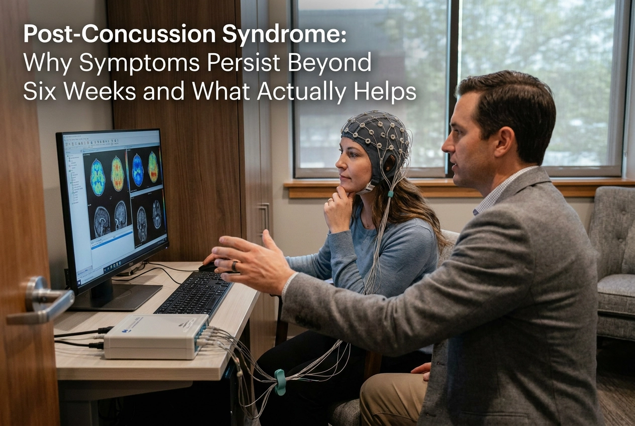
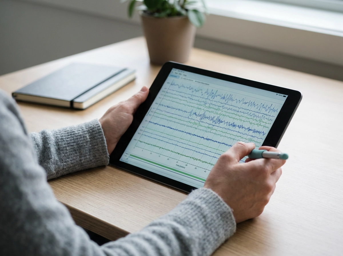
qEEG records the brain's electrical activity across multiple frequency bands simultaneously, including delta, theta, alpha, beta, and gamma waves, across dozens of scalp locations. The result is a detailed map showing which regions are generating abnormal patterns, which frequencies are running too fast or too slow, and how severe the dysregulation is compared to a normative database of healthy brains. It measures characteristics like amplitude, coherence, and phase relationships, giving clinicians a precise picture of how the brain is actually functioning at the time of assessment.
Why Structural Imaging Is Not Enough
A normal MRI or CT scan does not mean a normal brain. Those tools are designed to detect structural damage: bleeding, lesions, fractures. They are not built to capture the functional disruptions that define PCS. A 2024 peer-reviewed study published in NeuroRegulation found that qEEG significantly differentiated acute mild TBI patients from neurotypical controls using power spectral and connectivity analyses, reporting highly significant effect sizes. That differentiation is invisible on conventional imaging. For patients who have been told their scans look fine but still feel impaired, qEEG addresses exactly that gap.
From Diagnostic to Directional
qEEG data does not simply confirm that a problem exists; it identifies where it is and what form it takes. That specificity matters because neurofeedback protocols are not one-size-fits-all. The frequency patterns that need to be trained, and the brain regions being targeted, differ from patient to patient. Without a qEEG baseline, a clinician is working from symptoms alone, which describes what the patient experiences but not the underlying neural mechanism producing it. Treatment planning that starts with qEEG targets the mechanism, not just the complaint.
Neuron Connect's Assessment Approach
At Neuron Connect, qEEG brain mapping is the clinical starting point for every concussion recovery case. No generalized protocol is applied before the data is reviewed. This matters when choosing where to seek care; not all providers begin with measurement. If you are evaluating options, what to look for in a mental health clinic outlines the criteria that separate data-driven neuroscience care from generic approaches.
Documentation Value
For patients in personal injury or workers' compensation cases, qEEG also provides an objective, timestamped record of functional brain status. That documentation carries more evidentiary weight than symptom self-report and can support legal and insurance proceedings with measurable clinical data.
What to Expect from Neurofeedback for Concussion Recovery
Once qEEG mapping has identified which brainwave patterns need correction, neurofeedback is how the brain is trained to shift them.
The process is straightforward and entirely non-invasive. Sensors placed on the scalp read the brain's electrical activity in real time. That data is fed back to the patient through a visual display or audio signal, and the brain responds by adjusting its own activity to keep the feedback favorable. This is operant conditioning applied at the neurological level: the brain receives moment-to-moment information about what it is doing and gradually learns to produce healthier frequency patterns. There is no stimulation, no discomfort, and nothing the patient needs to consciously "do" beyond staying engaged with the screen or audio. Think of it as a workout for the neural pathways, where the feedback signal is the resistance that drives adaptation.
What Sessions Actually Look Like
Sessions are conducted in a clinical setting and typically run 30 to 60 minutes. The patient sits comfortably, sensors in place, and observes the feedback display. That is the full experience from the patient's perspective. The complexity happens in the software, which compares incoming brainwave data against the target parameters set by the clinician and adjusts the feedback signal accordingly throughout the session.
How Long Recovery Takes
Symptom changes are gradual, not immediate. Most protocols extend across multiple sessions over several weeks, and the total number of sessions is determined by the individual's qEEG profile and the severity of their symptom presentation. There is no universal session count because no two qEEG profiles are identical.
Patients commonly report early gains in sleep quality, emotional steadiness, or reduced headache frequency before more demanding functions improve. Sustained concentration and memory consolidation tend to show measurable progress later in the protocol, once foundational regulatory stability has been established.
Setting Realistic Expectations
Neurofeedback is a neurological rehabilitation process. Progress should be measured in functional terms: sleeping more consistently, tolerating cognitive demands longer, reacting less intensely to sensory input. Framing outcomes as measurable functional improvements, rather than complete symptom elimination, reflects both the current evidence base and the actual clinical experience of most patients.
Because the field carries formal clinical responsibility, practitioner credentials matter. The International Society for Neuroregulation and Research (ISNR) maintains certification standards and published practice guidelines specifically to ensure that neurofeedback for concussion recovery is delivered within established clinical parameters. Before beginning care, patients should confirm that their provider holds recognized credentials and that treatment is guided by qEEG data rather than a one-size-fits-all protocol.
When to Seek Specialized Help for Post-Concussion Syndrome
Knowing when to stop waiting and seek specialized care is often the hardest part of concussion recovery.
The clearest indicator is symptom persistence beyond six weeks despite following standard rest-based guidance. At that point, passive recovery has not worked. The nervous system has not self-corrected, and continuing the same approach is unlikely to produce a different result.
Several additional signals warrant prompt evaluation:
Symptoms that are worsening rather than plateauing
New symptoms appearing after an initial period of partial improvement
Meaningful functional impairment at work, in school, or in daily tasks that were previously manageable
Any of these patterns suggests the brain has stabilized around a dysregulated state that rest alone cannot resolve.
A normal MRI or CT result does not close the case. Structural imaging is designed to detect tissue damage, bleeds, and physical abnormalities. It cannot capture the brainwave dysregulation and altered connectivity that drive persistent post-concussion syndrome. Patients who have been told their scan is normal but remain symptomatic should specifically seek providers capable of functional neurological assessment. A clear scan rules out structural injury; it does not rule out PCS.
Referrals into specialized care can come from multiple directions. Primary care physicians, neurologists, and sports medicine providers are common entry points. Attorneys managing personal injury or workers' compensation cases may also refer clients when documented functional impairment is relevant to the legal record, since objective neurophysiological data carries more evidentiary weight than symptom self-report alone.
Arizona residents dealing with persistent symptoms after a concussion can take a concrete next step. Recovering after a concussion or head injury? Neuron Connect provides qEEG brain mapping consultations at locations serving Phoenix, Scottsdale, and Tucson. The assessment maps actual brainwave activity to determine whether measurable dysregulation is present and whether active neurological rehabilitation is appropriate for that specific presentation. It replaces guesswork with data, and waiting with a documented plan.
Takeaways: Moving from Passive Waiting to Active Recovery
The evidence reviewed throughout this post points to a single, clear conclusion: post-concussion syndrome is not slow healing. It is a measurable neurophysiological condition, sustained by brainwave dysregulation and altered neural connectivity, and it requires a fundamentally different response than rest and time.
Here is what that means in practice:
Rest serves a purpose acutely, reducing symptom load in the days immediately following injury. Beyond six weeks, however, rest alone cannot correct dysregulated frequency patterns or restore desynchronized communication between brain regions. Waiting longer does not change the underlying mechanism.
Measurement comes before treatment. qEEG brain mapping identifies precisely which frequencies are dysregulated, in which regions, and to what degree. That data is what makes neurofeedback and cognitive rehabilitation therapy for TBI genuinely targeted rather than generalized. Without it, treatment is an educated guess.
Active rehabilitation has a documented clinical pathway. Institutions including the International Society for Neuroregulation and systematic review bodies have formally evaluated qEEG-guided neurofeedback for concussion recovery. The evidence base is growing, the professional standards exist, and the interventions are available now.
A normal MRI does not mean nothing is wrong. Structural imaging misses functional disruption entirely. Patients who have been told their scans look fine and are still symptomatic have not exhausted their options; they have not yet had the right assessment.
If you or someone you care for is still struggling weeks or months after a concussion, more time on the couch is not the answer. The next step is an objective look at what is actually happening in the brain.
At Neuron Connect, qEEG brain mapping is the starting point for every concussion recovery case. Arizona residents in Phoenix, Scottsdale, and Tucson can schedule a consultation to find out whether active neurological rehabilitation is the appropriate next step for their specific situation.
Conclusion
Post-concussion syndrome is not a waiting game. The brain has a measurable, addressable problem, and passive rest simply does not resolve functional disruption at the neurological level. What does help is objective assessment through qEEG brain mapping, followed by targeted active rehabilitation protocols that address the actual source of persistent symptoms.
Three realities define better outcomes: symptoms beyond six weeks signal a specific clinical condition, not slow healing; structural scans miss what functional assessments catch; and evidence-based treatment exists right now for those willing to pursue it.
If you are still suffering weeks or months after a concussion, you deserve answers grounded in data, not reassurance based on a normal MRI. Take the next concrete step. Schedule a qEEG brain mapping consultation at Neuron Connect and find out exactly what your brain needs to recover.